|
|
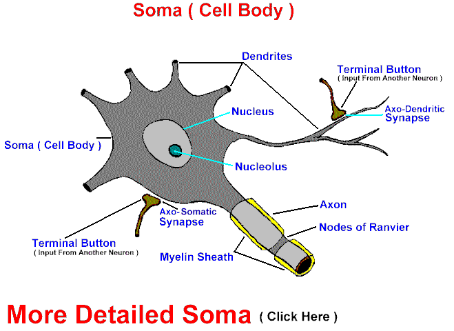
The surfaces of most cell bodies have receptor sites for numerous nerve endings. The cytoplasm contains the usual organelles found in cells, such as mitochondria, lysosomes, and microtubules. Some neurons may contain such large quantities of melanin that they form nuclei (such as the substantia nigra) with grossly much darker pigmentation than the surrounding tissue. Also, with aging, lipofuscin granules can accumulate to occupy half of the cytoplasm ( neurons never divide and last a lifetime hopefully ). Well developed polyribosomes and rough endoplasmic reticulum may be visible with light microscopy as basophilic patches, called Nissl substance. This is easily seen in large active neurons such as the motor neurons of the spinal cord, and poorly visible in small sensory neurons. Abundant, 10 nM thick intermediate filaments, or neurofilaments, help form the cytoskeleton of the perikaryon.
Material constantly travels from the cell body down the axon, called anterograde flow; microtubules are thought to play a role in some of the movement. A slow flow component, moving at 1-4.5 mm/day, is seen with neurotransmitters, hormones and protein for membrane renewal. This is also the maximal rate of regeneration of nerve fibers following injury. Amino acids, polypeptides and small proteins can move much faster, up to 400mm/day.
|